

Quick Answer
A CBCT (cone beam computed tomography) scan uses a rotating X-ray beam to capture hundreds of images from different angles, which are combined into a detailed 3D model of your teeth, jawbone, nerves, and sinuses. It takes under a minute and is painless.
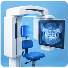
How CBCT Differs from a Regular Dental X-Ray
Standard X-rays are 2D: Traditional dental X-rays (bitewings, periapicals, panoramic) produce flat, two-dimensional images. They are excellent for detecting cavities, bone levels, and tooth roots, but they cannot show depth.
CBCT produces a 3D model: A CBCT scanner rotates around your head, capturing 150–600 individual images from different angles. Software combines these into a full 3D reconstruction that can be viewed from any angle and in any cross-section.
More detail, more information: CBCT reveals the exact position of nerves, the volume and density of bone, the relationship between teeth and anatomical structures, and the precise location of pathology that 2D X-rays may miss.
When Is a CBCT Scan Used?
Implant planning: The most common use. CBCT measures bone height, width, and density at the implant site and identifies the exact location of the inferior alveolar nerve to prevent injury during surgery.
Wisdom tooth evaluation: When a wisdom tooth is close to the inferior alveolar nerve, a CBCT scan shows the precise relationship and helps the surgeon plan a safer extraction.
Bone graft assessment: Before and after bone grafting, CBCT confirms whether sufficient bone volume has been achieved for implant placement.
Complex root canals: CBCT reveals extra canals, root fractures, and the 3D anatomy of root systems that are difficult to see on standard X-rays.
TMJ evaluation: CBCT provides detailed images of the temporomandibular joint to assess bone changes, joint space, and condyle position.
Frequently Asked Questions
Questions About Your Imaging?
Our team is happy to explain any imaging we recommend and why it is needed for your treatment. Schedule a consultation today.